
*The primary advantage of the paralleling
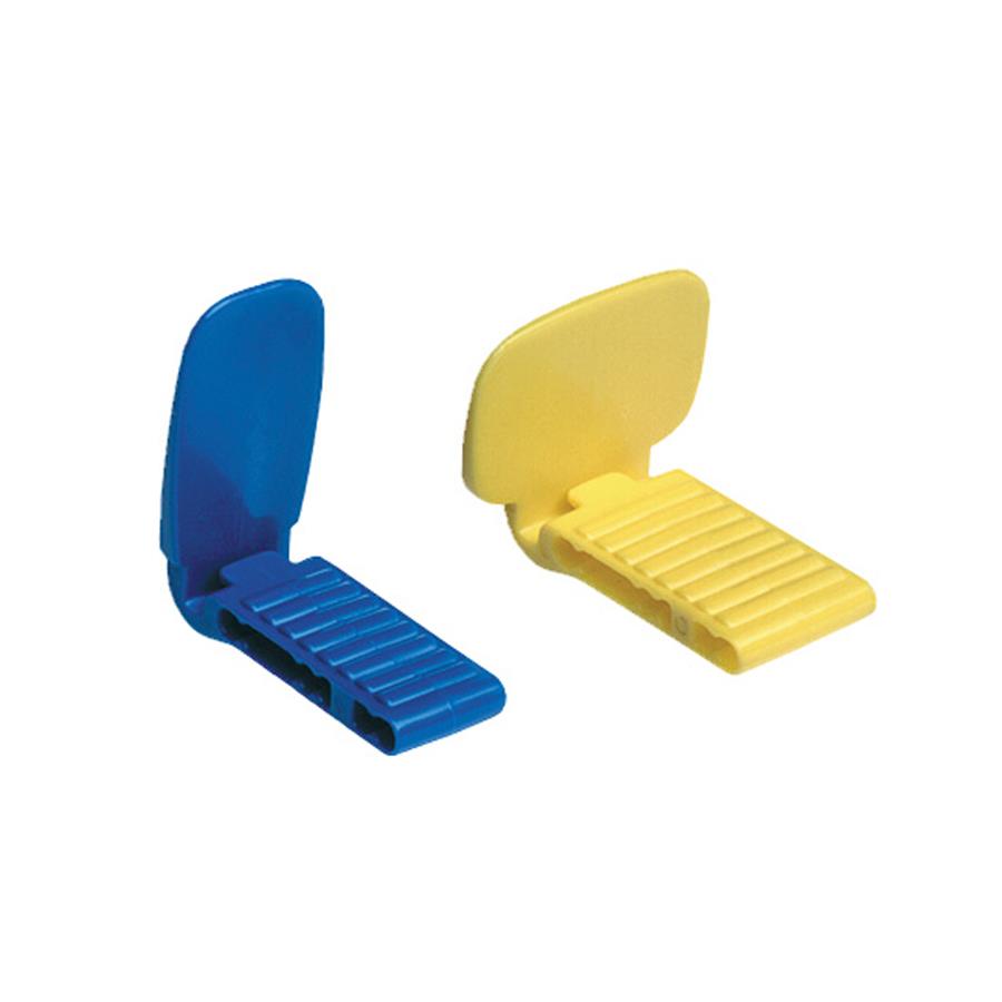
STABE BITE BLOCK SERIAL
Comparison of serial images has great validity. It eliminates chances of dimensional distortion. It eliminates the need to determine horizontal and The receptor must be placed between the tori and the tongue The receptor must be placed on the far side of the torus and then The vertical angulation can be increased by 5 to 15 degrees. Two cotton rolls can be used, one placed on each side of the Taking a dental image of diagnostic quality. Placement will assist the radiographer in Using cotton rolls and changing the verticalĪngulation and the location of the receptor

To be competent in dealing with these types of
STABE BITE BLOCK SERIES
Specific placements described in the chapter areįor 15-receptor periapical series using size 1 Dictated by teeth and surrounding structures The specific area where the receptor must be.Premolar receptor first and then the molar receptor?24 In each quadrant, why should you always expose the Make this sequence a habit for all imaging exposures. Stick to a sequence: Do not interchange from patient Four maxillary exposures four mandibular exposures Size 1 receptor is small and easier for patient to tolerate Anterior exposure sequence (always start with the anterior teeth).Open the sterilized package containing the Receptor placement for paralleling technique Exposure sequence for receptor placements The x-ray beam must be centered on the receptor to The central ray of the x-ray beam must be directed through he Perpendicular to the receptor and long axis of the Central ray of the x-ray beam must be directed Receptor must be positioned parallel to the long axis Receptor must be positioned to cover the prescribed Long portion in the horizontal direction Rinn XCP-ORA One Ring & Arm Positioning System Rinn XCP Extension Cone Paralleling System Examples of commercially available intraoral.Mouth and retain the receptor in position A device used to position the receptor in the.Most parallel rays will be directed at the tooth Must be increased to ensure that only the Keep the receptor parallel with the long axis of Perpendicular to the film and the long axis of The central ray of the x-ray beam is directed The long axis of the tooth being radiographed. The receptor is placed in the mouth parallel to.To expose periapical and bite-wing image receptors. Paralleling technique is one method that can be used Paralleling technique is also known as:.Placement procedures used in the paralleling Preparation, equipment preparation, and receptor To present basic concepts and to describe patient List the advantages and disadvantages of the That are used for a patient with a shallow palate, bonyġ3. Explain the modifications in the paralleling technique Summarize the guidelines for periapical receptorġ2. Recommended for use with the Rinn XCP instruments. Receptor placements using the paralleling technique ĭescribe each of the 15 periapical receptor placements Discuss the exposure sequence for 15 periapical Describe the patient and equipment preparations thatĪre necessary before using the paralleling technique.ġ0. State the five basic rules of the paralleling technique.ĩ. Paralleling technique and how each receptor is placed

Describe the different sizes of receptors used with the Identify and label the parts of the Rinn XCPħ. List the beam alignment devices that can be used withĦ. Describe why a beam alignment device is necessaryĥ. Discuss how object-receptor distance affects the imageĪnd how target-receptor distance is used to

State the basic principle of the paralleling techniqueĪnd illustrate the placement of the receptor, beamĪlignment device, position-indicating device (PID), andģ. Define the key terms associated with the parallelingĢ.


 0 kommentar(er)
